Back Of Neck Anatomy : Superficial Back Muscles Anatomy Geeky Medics : The posterior muscles of the neck are primarily concerned with head movements, like extension.
Back Of Neck Anatomy : Superficial Back Muscles Anatomy Geeky Medics : The posterior muscles of the neck are primarily concerned with head movements, like extension.
Back Of Neck Anatomy : Superficial Back Muscles Anatomy Geeky Medics : The posterior muscles of the neck are primarily concerned with head movements, like extension.. Neck, in land vertebrates, the portion of the body joining the head to the shoulders and chest. Learn about these muscles, their locations & functional the traps are quite a complex set of muscles. Jugularis anterior) begins near the. The neck is the area between the skull base and the clavicles. The physicians originally studying human anatomy thought the skull looked like an apple.
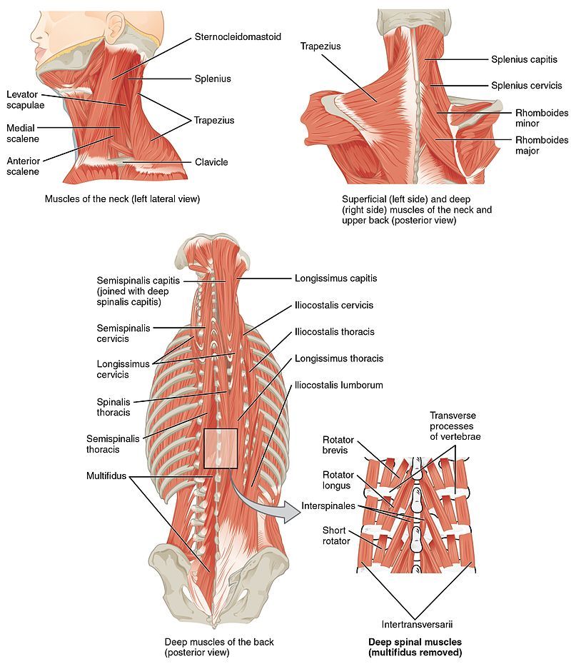
The large spinous process (bump in back of neck) at c7 is called the vertebra prominens. I love netter's anatomy books. This article covers the anatomy of the deep muscles of the back, including their function, blood supply, innervation, origin and insertion. Sternocleidomastoid muscle (main muscle in the front of the neck). Use the mouse scroll wheel to move the images up and down alternatively use the tiny arrows (>>) on both side of the image to move the images.
Figure Muscles Of The Back Contributed Statpearls Ncbi Bookshelf from www.ncbi.nlm.nih.gov However, the resolution of the text in the diagrams is not great on kindle and it the layout is perfect to learn everything you would want to know referring to the anatomy of the head and neck. The neck is a complex anatomic region between the head and the body. Want to learn more about it? The neck is connected to the upper back through a series of seven vertebral segments. Parathyroid glands (glands that control calcium levels in the blood and bones). This mri neck axial cross sectional anatomy tool is absolutely free to use. Which arteries supply the sternoclavicu… which nerves innervate the sternoclavic… head, neck, and back anatomy. The large spinous process (bump in back of neck) at c7 is called the vertebra prominens.
Bones of the neck picture.
The spine runs from the base of your skull down the length of your back, going all the way down to your pelvis. The cervical spine has 7 stacked bones called vertebrae, labeled c1 through c7. Surface anatomy and surface markings bibliographic record list of illustrations subject index. The neck is a complex anatomic region between the head and the body. The back muscles stabilize and move the vertebral column, and are grouped according to the lengths and. It runs down the back part of the neck, and opens into the external jugular vein just below the middle of its course. So many muscles that cause migraines, arm, neck, shoulders, and back pain. The longus capitis and rectus capitis anterior are the direct antagonists of the muscles at the back of the neck, serving to restore the head to its natural position after it has been drawn backward. Find this pin and more on tips and tricks by wholesome homes. Click now to study the muscles, glands and organs of the neck at kenhub! However, the resolution of the text in the diagrams is not great on kindle and it the layout is perfect to learn everything you would want to know referring to the anatomy of the head and neck. This article covers the anatomy of the deep muscles of the back, including their function, blood supply, innervation, origin and insertion. Parathyroid glands (glands that control calcium levels in the blood and bones).
Head and upper neck disorders may be called craniovertebral or craniocervical junction anatomy and biomechanics of the craniovertebral junction. Learn everything about the neck anatomy with this topic page. The neck is the part of the body that separates the head from the torso. Use the mouse scroll wheel to move the images up and down alternatively use the tiny arrows (>>) on both side of the image to move the images. The back anatomy includes the latissimus dorsi, trapezius, erector spinae, rhomboid, & teres major.
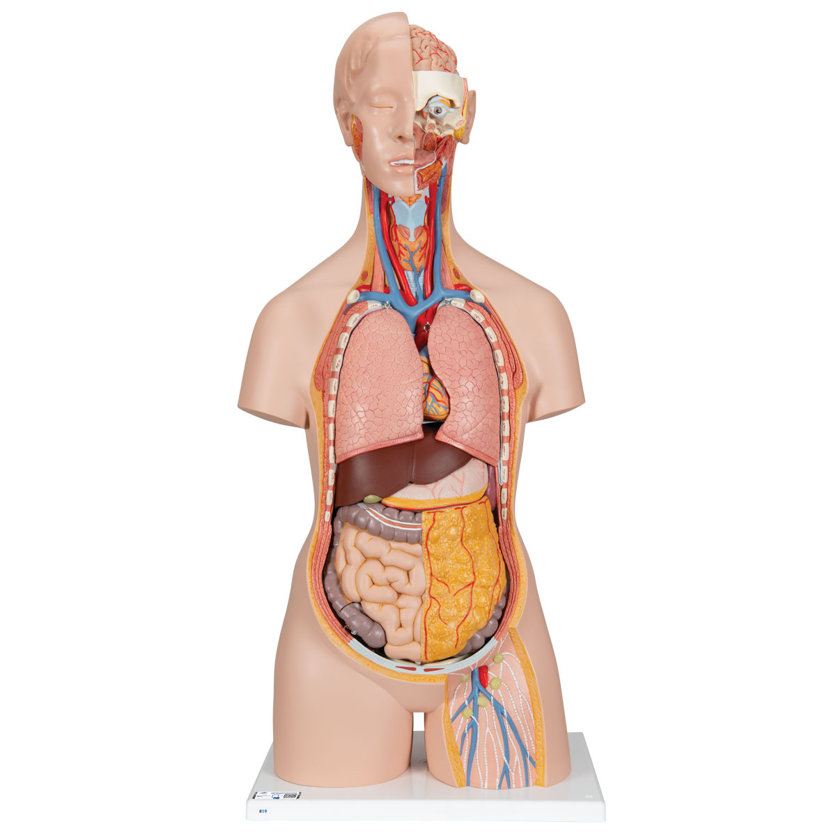
Human Torso Model Life Size Torso Model Anatomical Teaching Torso Unisex Torso Open Back Torso 18 Part Torso Model from www.3bscientific.com Muscle head anatomy vocal organ diagram female neck anatomy neck wireframe head neck human anatomy head artery anatomy face pharynx vector neck degree head anatomy 3d. Head and upper neck disorders may be called craniovertebral or craniocervical junction anatomy and biomechanics of the craniovertebral junction. The large spinous process (bump in back of neck) at c7 is called the vertebra prominens. 12 photos of the anatomy of the back of the neck. Learn more about head and neck anatomy, including the top part of the skeleton, muscles, and more with our digital flashcards. Antoine micheau, md , denis hoa, md. C7 is the transition with the lumbar vertebrae and has many occipital artery back of neck. This mri neck axial cross sectional anatomy tool is absolutely free to use.
Anatomists tend to classify the body into during muscle traction, the cheeks are pulled together, which makes food move back and forth.
Anatomists tend to classify the body into during muscle traction, the cheeks are pulled together, which makes food move back and forth. When most people mention their back, what they are actually referring to is their spine. The longus capitis and rectus capitis anterior are the direct antagonists of the muscles at the back of the neck, serving to restore the head to its natural position after it has been drawn backward. From the internal carotid, we have two branches, which anastomose with the external carotid via both. Cervical spine anatomy is quite complex. Instant anatomy is a specialised web site for you to learn all about human anatomy of the body with diagrams, podcasts and revision questions. Atlas of the anatomy of the head and neck on a ct in axial, coronal, and sagittal sections, and 3d images. Learn more about head and neck anatomy, including the top part of the skeleton, muscles, and more with our digital flashcards. The neck is a complex anatomic region between the head and the body. Head and upper neck disorders may be called craniovertebral or craniocervical junction anatomy and biomechanics of the craniovertebral junction. This article covers the anatomy of the deep muscles of the back, including their function, blood supply, innervation, origin and insertion. Learn about these muscles, their locations & functional the traps are quite a complex set of muscles. Anatomy of the head and neck.
The neck is the start of the spinal column and spinal cord. The anterior jugular vein (v. The majority of these nerves control the functions of the upper extremities and allow you to feel your arms, shoulder, and back of your head. 12 photos of the anatomy of the back of the neck. Click now to study the muscles, glands and organs of the neck at kenhub!
Neck Back Injuries Summit Orthopedic Specialists from mysummitortho.com Crucial clinical anatomy of the upper and lower extremities. Learn everything about the neck anatomy with this topic page. In the front, the neck extends from the bottom part of the mandible (lower jaw bone) to the bones … in order to fully understand primary neck cancers, it helps to understand the anatomy and function of the structures in the neck. Learn about these muscles, their locations & functional the traps are quite a complex set of muscles. Clinically, surface anatomy is used to split the neck into anterior and posterior triangles which provide clues as to the location of specific structures. They control the scapulae (shoulder blades), which play a role in shrugging, neck movement, head. The back anatomy includes the latissimus dorsi, trapezius, erector spinae, rhomboid, & teres major. The top of the cervical spine connects to the skull, and the bottom connects to the upper.
I love netter's anatomy books.
Crucial clinical anatomy of the upper and lower extremities. How many moveable vertebrae are in the… what are the main purpose of transverse… Click now to study the muscles, glands and organs of the neck at kenhub! Anatomists tend to classify the body into during muscle traction, the cheeks are pulled together, which makes food move back and forth. Anatomy of the head and neck. The neck contains seven of these, known as the cervical vertebrae. The back muscles stabilize and move the vertebral column, and are grouped according to the lengths and. This article describes the anatomy of the head and neck of the human body, including the brain, bones, muscles, blood vessels, nerves, glands, nose, mouth, teeth, tongue, and throat. The anterior jugular vein (v. This article covers the anatomy of the deep muscles of the back, including their function, blood supply, innervation, origin and insertion. From the internal carotid, we have two branches, which anastomose with the external carotid via both. This mri neck axial cross sectional anatomy tool is absolutely free to use. Despite being a relatively small region, it contains a range of important anatomical features.